Sure Developers Team Services

Sure Developers Team Services

Translate

Barrett's oesophagus

Barrett's oesophagus


Barrett's oesophagus


BARRETT'S OESOPHAGUS= also known as columnar lined oesophagus.    Def-whn metaplastic change occur in lining mucosa of oesophagus, thn it is called barrett's oesophagus.. It is premalingnant lesion n ulcer in barrett's clo is barrett's ulcer.   B)PATHOGENESIS=  repeated reflux results in shifting of oesephagogastric junction upwards wich increases reflux resultimg in intestinal metaplasia of middle n lower oesophagus.  C)pathological types= 1- gastric type wid chief n parietal cells. 2-intestinal type..3- junbtional type.. D)clinical types..= 1-long segment..2-short segment.. E)incidence of malignancy= 25 times more prone to CA lower n middle oesophagus.. F)types of dysphasia= 1-low grade..2-high grade.. G)carcinoma in barrett's oesophagus= 1- it wil b invasive..2- it is more proximal.. 3- carries poor prognosis. 4- 25 times increased risk.  H) TREATMENT= 1-laser photodynamic therapy..2-argon beam plasma coagulation..3- high dose proton pump inhibitors for 8 wks. 4- oesophagectomy in cases of high grade dysplasia.. I)COMPLICATIONS= 1- oesophageal ulber..2-oesophageal stricture..3-dysplasia ..4-adenocarcinoma.. 

Benign ovarian tumour

Benign ovarian tumour


Benign ovarian tumour

Benign ovarian tumour-
80%of ovarian tumour are benign.They can become secondary malignant however.
C/O-
symptoms_1 abdominal swelling upto 50 kg ovarian tomour hv been noted. 2 irregular menstrual cycle-theca granulosa cell tumour_oestrogen secretion which cause menorrhagia..
Musculizing tumour_amenorrhoe and virilization.
Brenner tumour_postmemopausal bleeding.
3 pressure symptoms-frequency of micturation,retention,pressure on rectum,dyspnoa,palpitation,bialateral pitting oedema of foot.
4 normally there is no pain but acute abdominal pain may occure if ovarian tumour undergoes torsion or rupture.
Physical sign-
inrpectiรถn-abdominal swelling formed by abdominal cyst.Abdominal wall moves with deep inspiration.Symmetrical position of tumour in abdomen.
Palpation-upper and lateral limit of tumour can b defind.Sooth surface. Small cyst.,tense and cystic.Large tumour is fixed. Fluid thrill can b illiciated.
Percussion-
dull over centre of tumour.Resonat over flank.
Auscultation-silent
 Bimannual examinatiรถn-
small tumour-uterus can b identified without difficulty.Cyst usually displace uterus to opposite side..If there is hard nodule it indicate malignancy.
D/D-
1 full bladder. 2 pregnant uterus
3 myoma 4 ascites
investigation-
1 Radiograph of abdomen and pelvis-soft tissue shadow,teeth in dermoid.
2 Diagnostic laparoscopic examination,iv pyelography excledes hydronephrosis.
3 Breast examinatiรถn to exclue pregnancy.
4 ultrasรถnograqy.
Benign cyst shows these features-unilateral,unilocular/multilocular with thin wall non-echogenic cavity,tumour marker CA 125 <35 unite/ml.
5 colour flow doppler-
neovascularization in malignant form.
6 CT MRI to identify dermoid cyst,haemorrhage cyst.
7 tissue marker CA 125 >35 unit/ml
CEA >5 mg/lit in mucinous ovarian tumour.
8 cytological study of ascitic fluid or aspirated cystic fluid.
Treatement-
laparotomy is needed.
Abdominal hysterctomy and bilateral salpingo-oophorectomy in perimenausal women.
Histological specimen may discover microscopic evidence of malignancy and thereby need of secondary surgery is avoided.
Ovarioctomy/cystectomy-
conserve healthy overy in young pt.
Clamp infudibulo pelvic ligament laterally mesovarian in middle,fallopian tuae ,overian liagment medially.
Laperoscopic cystectomy/ovariotomy_ minimal invasive surgery in vague for small cyst.
First aspirate cystic fluid,then dissect cyst wall.

Bact. Meningitis

Bact. Meningitis
Bact. Meningitis


Bact. Meningitis
Acute bacterial meningitis a major cause of morbidity and mortality in young children occurs both in epidemic and sporadic pattern.
Etiopathogenesis
The common organisms implicated in neonates are E.coli,steptococcus pneumoniae,salmonella species,pseudomonas aeruginosa,streptococcus fecalis and staph aureus.from age of 3 months to 3 yrs infection is due to hemophilus influenzae,s pneumoniae and meningococci.beyond 3 yrs 2 most common organisms r pneumococcus n meningococcus.
The infection usually spreads hematogenously to meninges from the distant foci of sepsis such as pneumonia, empyema,pyoderma and ostemyelitis.purulent meningitis may follow head injury.rarely the infection may extend to the meninges from contagious septic focus in the skull and spine.eg.infected papa nasal sinuses,mastoiditis,osteomyelitis, and fracture of the case of skull.
Recurrence may be associated with pilonidal sinus,CSF rhinorrhea,traumatic lesions of cribriform plate and ethmoidal sinus or cong. fistulae.

Clinical features
The onset is usually acute and febrile.in the initial phase the child becomes irritable resents light has bursting headache either diffuse or in the frontal region,spreading to the neck n eyeballs.the infant may have projectile vomiting,shrill cry and a bulging fontanel.
Seizures r a common symptom n may occur at the onset or during the course of illness.varying grades of alterations in sensoriun may occur.photo phobia is marked.there is generalised hyper tonia and marked neck rigidity.flexion of the neck is painful n limited.kernigs sign is present.there may b pain in the back of the thigh or muscles of the back.in brudzinski sign knees get flexed as neck of the child is passively flexed.the fundus is either normal or shows congestion and papilledema.extrinsic ocular palsies may lead to squint diplopia n ptosis.he skin of abdomen is lightly scratched flush may be seen.the muscle power in the limbs are preserved.reflexes are normal diminished or exaggerated.respiration may become periodic or cheyne stokes type often with shock.

Lab diagnosis
1) History of fever irritability photo phobia headache vomiting convulsions and altered sensorium.
2) CSF exam; it has elevated pressure.it is turbid wid an elevated cell count >1000/mm3 and mostly polymorphonuclear.proteins r elevated above 100mg/dl and sugar reduced to below 50%.
3) CT Scan useful to exclude sub dura effusion.brain abscess, hydrocephalus,exudates and vascular complications.
4) rapid diagnostic tests distinguish between bacterial viral and tuberculous meningitis based on antigen n antibody demo.eg. Latex agglu. Immune electrophoresis. ELISA.
5) polymerase chain reaction 4 viral m.
6) non specific tests like C reactive proteins,lactic dehydrogenase.

Complications
Subdural effusion or empyema.
Ventriculitis
Arachnoiditis
Brain abscess
Hydrocephalus.

Treatment
Initial empiric treatment
It should begin with one of third gen.cephlosporins such as cefotaxime or ceftriaxone.a combination of ampicillin 200mg/kg and chloramphenicol 100/mg/kg/day for 10 to 14 days.
Specific therapy
1)for meningococcal or pneumococcal meningitis.penicillin 4-5 lac units/kg/day 4 hrly.ceotaxime(200mg/kg/day) or ceftriaxone(150mg/kg/day) also effect.
2)H. Influenzae meni.ceftriaxone or cefo i.v. Used as single agent.
3) staph meni. Vancomycin is Rx of choice if peni. Or methi. resistance suspected
4) listeria. Ampicillin 300mg/kg/day and aminoglycoside prefer.
5)pseudomonas.combination of ceftazidime n aminogl. Is used.

Steroid therapy: dexamethasone in a dose of 0.15 mg/kg iv 6 hrly for 5 days.

DDs
Meningism
Partially treated bacterial meningitis.
Aseptic m.
Tuberculous m.
Cryptococcal m.
Viral encephalitis
Poliomyelitis
Subarachnoid hemorrhage.
Lyme disease

BCG

BCG
BCG


BCG


It is an attenuated strain of mycobacterium tuberculosis var bovis used as a live vaccine against tb.

1 Dose
       0.1ml i.d
2 site
       left deltoid
3 schedule
       at birth
4 Reaction
      5mm induration -
            !
        papule
            ! 2-3wk
        pistule-
            !
         ulcer
            ! 6-12wk
         scar
5 heat labile

6 light sensative

7 store
       at 2-8'c
8 SIDE EFFECT
        1 LOCAL BACT INFECTION
        2 KELoid
9 advantage
       prevent  1 neuro tb
                 2 miliary tb
                 3 hemato tb

10 infants became positive for           tuberculin test after 4 to 12          weeks of immunization

11

Bell's palsy

Bell's palsy
Bell's palsy


Bell's palsy

Aetiology
herpes simplex virus and herpes zoster
PATHOLOGICALLY
odema and swelling of the nerve accur witho facial canal ,often at stylmastoid foramen
CLINICAL FEATURES
FEATURES OF LMN PALSY
1 PARALSIS OF ALL THE MUSCLES OF ONE SIDE OF THE FACE
2.Drooping of the palsy ,effacement of creases and skin dole on affected side .
3.weakness of frowning and eyes closures the upper facial muscles are weak .
4. Drooling of saliva from angle of mouth .
5. On asking the patient to show his teeth ,the angle of the mouth deviates away from the side of lesion
6.upon attempted closure of the eyelid .
the eye on the paralysed side rolls upwards .
7. Corneal ulceration due to inability to close the eye during sleep.
,
INVESTIGATION
1. ELECTROPHYSIOLOGICAL test
2. To rule out alternate diagnosis , appropriate diagnostic procedures may be necessary.
,,
TREATMENT
PREDNISOLONE AND ACYCLOVIR
-Prednisolon 1mg/kg /day for 1st wits the dosage tapering off over the 2nd week.
- Aciclovir at a dosage of 800mg five times a day for 5days.   ,
  

Baby friendly hospital

Baby friendly hospital

Baby friendly hospital

The program was launched in 1992 by UNICEF & WHO with an abjective to reestablish the superiority of breast feeding in order to protect the newborn's health by becoming baby friendly.

Following steps are included:
1. Written breast feeding policy
2. Entire staff must be trained to implement the policy
3. Inform all pregnant women about benefits of breast feeding
4. Mothers should be helped to initiate breast feeding within half hour of birth
5. Mothers are shown the best way to breast feed.
6. Unless medically indicated, the newborn should be given no other food or drink other than breast milk.
7. To practice 'rooming in' by allowing mothers & babies to remain together for 24 hours.
8. To encourage demand breast feeding.
9. No artificial teats should be given.
10. Breast feeding support groups established & mothers referred to them on discharge.
BEST OF LUCK DUDE.. By Abhishek

Acute osteomyelitis

Acute osteomyelitis
Acute osteomyelitis

Acute osteomyelitis

Etiopaho-
1 staph.aureus
2 strepto
3 pneumcoccus
paho
host bone initiates an infamatory reaction in responce to bacteria.This leads to bone destruction n production of an inflamatory exudate n cels.
Pus spreads in the folowing dir
a)Along the medulary cavity-

Acromegaly

Acromegaly
Acromegaly

Acromegaly

AETIOLOGY-
1. GH hypersecretion in adult life after epiphyseal closure result in acromegaly
2.comman - pituitary tumor
   rare - pancreatic islet tumor,
          bronchial carcinoid,
         small cell lung carcinoma

CLINICAL FEATURE -
1. Soft tissue changes -
    -thickening of skin
-increased skin tags

Active management of 3rd stage of labour

Active management of 3rd stage of labour

Active management of 3rd stage of labour

THE UNDERLYING PRINCIPLE IN ACTIVE MANAGEMENT =it is to excite powerful uterine contractions following birth of anterior shoulder by parenteral oxytocin which facilitates not only early seperation of placenta but produces effective uterine contractions following its seperation.


Acute bronchiolitis

Acute bronchiolitis
Acute bronchiolitis

ACUTE BRONCHIOLITIS:

 IT is 1 of the common serious acute lower respiratory infections in infants.
- age bet^ 1 to 6 mnt.
- common in winter n spring.
- respirtory syncytial virus is common couse. Other r Parainfluenza virus 3,1 n 2, adenovirus, influenza.

PATHOGENESIS

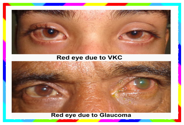
Acute congestive glaucoma

Acute congestive glaucoma

Acute congestive glaucoma

An attack of acute primary angle closure glaucoma occurs due to a sudden total angle closure leading to severe rise in IOP.

Clinical features
Symptoms
- pain: sudden รถnset of very severe pain
- nausea, vomiting and prostrations

Acute dacrocystitis

Acute dacrocystitis
Acute dacrocystitis

ACUTE DACRYOCYSTITIS...

 It is an acute suppurative inflammation of lacrimal sac , characterised by presence of painful swelling in the region of sac..
ETIOLOGY,
1. As an acute exacerbation of chronic dacryocystitis..
2. As an acute peridacryocystitis due to direct involvement from the neighbouring infected structures ..

Acute glomerulonephritis

Acute glomerulonephritis

Acute glomerulonephritis

 it is glomerular inflammation in which there is an immunological mediated injury to the glomeruli
AETIOLOGY
1.INFECTIOUS DISEASE
post streptococcal glomerulonephritis, infective endocarditis ,syphilis , mumps,nearles
hepatitis b ,infectious mono nucleosis,epstein barr virus .
2.MULTI SYSTEM DISEASE

Acute myocardial infarction

  
Acute myocardial infarction

Acute myocardial infarction

 - Myocardial infarction is myocardial necrosis occuring as a result of a critical imbalance betn coronary blood supply and myocardial demand
   - Usualy due to atheromatous thrombus in coronary artery.
   - Hypercoagulable state in youngs result in MI
   - Use of cocain is another cause of MI. Cocain produces
hypercoagulable state an dvasospasm of coronay arteries

Acute pancratitis

Acute pancratitis
Acute pancratitis

Acute pancratitis

Etiology
 Alcohol ingestion
 Biliary calculus
 Post operative
 Post ERCP
 Trauma to abdomen
 Metabolic  renal failure, hyprcal
 Penetrating peptic ulcr
 Connective tissue disease SLE
 polyarteritis nodasa
 Infections mumps,roundwrm ,vir
  hepatitis
 Drugs sulphonamds
,oestrogen,thiazide ,frusemide,steroids.

Acute peritonitis

Acute peritonitis
Acute peritonitis

Acute peritonitis

Def=inflammation of peritoneum is called peritonitis

CAUSES
A)PRIMARY PERITONITIS
1)spontaneous peritonitis of childhood
2)spontaneous peritonitis of adults
3)tuberculous peritonitis
4)peritonitis a/w dialysis

B)SECONDARY PERITONITIS
peritonitis due to intra abdominal
source

Acute pancreatis

Acute pancreatis

Acute pancreatis

Causes
1>alcohol-it stimulate pancreatic secretion rich in protein,form protein plug,result in obstruction 2 pancretic duct.It stimulates trypsiogen
2>biliary tract disease-stone in biliary tree
3>collagen vascular disorder-polyarteritis nodosa
4>drug-corticosteroid,estrogen,diuretics
5>endoscopic procedure-sphicterectomy,cannulation of CBD or pancreatic duct or basketing of stones from CBD

Acute renal failure

Acute renal failure
Acute renal failure

Acute renal failure

DEF.

Rapid Reversible deterioration in renal function sufficient to result in accumulation of nitrogenous waste in body.

ETIOPATHOLOGY.

1 At rest 25% of co to kidney

2 in prerenal :- inadequate perfusion lead to decrease GFR lead to oliguria

ADAMANTINOMA

  
ADAMANTINOMA
ADAMANTINOMA

ADAMANTINOMA

Arises from ameloblasts
  Benign tumour, grows slowly, behaves like a basal cell carcinoma.
   Inadequate Rx results in local recurrence & metastasis. Hence Rx like malignant tumour
    * SITES
  mandible ( most common) , tibia, pgtuitary
    * C/f
- 4th or 5th decade
- it undergoes cystic degeneration
 resulting in multiple cystic spaces.Hence called multilocular cystic disease

Addisons disease

Addisons disease
Addisons disease

Addisons disease

Addisons disease=  primary causes        1 addisons disease
 2 congenital or acquired enzyme defects                        secondary causes  1. hypothalamic or pituitary 2.witgdrawal of glucocorticoid therapy.
Addisons disease-
causes-
1.Autoimmune adrenalitis-infectious adrenalitis,TB,fungal
2.Adrenal haemorrhage:-waterhouse-friderichsen syndrome,anticoagulation therapy,trauma.

Adenomyosis

Adenomyosis
Adenomyosis

Adenomyosis

*Def:
Adenomyosis is condition characterized by the presence of ectopic endometrial tissue  within the myometrium..
The condition is typically found in women between the ages of 35 and 50..
The disease coexist with uterine  fibromyoma and endomdtrial carcinoma.

*Grossly :
 uterus appears symmetricaly enlarged to not more than 14 weeks size.

ADHD

ADHD
ADHD

ATTENTION DEFICIT HYPERACTIVITY DISORDER

It is defined cp age inappropriate hyperactivity ,impulsiveness , and inattention .
It is divide into three classes
class1, all three symptoms
class2 first two only
class3 mainly the third

DIAGNOSIS
diagnosis is mainly based on clinical symptoms
INATTENTION :

Alcoholic liver disease

Alcoholic liver disease

Alcoholic liver disease

Etiopathogenesis=
.Alcohol intake..
Alcohol is metabolised by liver to acetaldehyde by mitochondrial enzyme,ADH.
Acetaldehyde forms adducts with cellular proteins in hepatocytes which activate immune system,leading to cell injury.
Acetaldehyde then metabolised to acetyl coA nd acetate by ALD,this generate NADH,which changes redox pontential of cell

Alopecia

Alopecia
Alopecia

Alopecia

It mean loss of hair n is a sign rather than diagnosis.
 - detail history, cair ful scalp exam^ n complete physical exam^ shud enable a confident diagnosis to b made.

CLASSIFICATION
LOCALISED
1 Non scarring
 - Tinea capitis
 - alopecia areata
 - androgenetic alopecia

Alzheimers disease

Alzheimers disease
Alzheimers disease

Alzheimers disease

Is a progressive neurodegenerative disease resulting in decline in congnitive functions and behavioural disturbances.It is the most common cause of cognitive impairment in elderly persons

c/f:

1. Insidious onset disease

2. There is a gradual decline in activities of daily living which ultimetly leads to profound disability and depandance on others

Amenorrhia

Amenorrhia
Amenorrhia

Amenorrhoea :

absence of menstruatino two types Primary secondary
primary amenorrhoea :failure of onset of menstruatino beyond age of 16 years regardless of development of secondary sexual character .
Classification it is made on basis of serum FSHlevel
! Hyper gonadotropic 2 eu gonadotropic 3 hypo gonadotropic
etiology: 1delayed puberty 2 cerebral stress emotional

AML

AML
AML

AML

OAML also termed as acute non lymphoblastic leukemia accounts for 15-20% of childhood leukemia.AML is much more complex and resistant disease than acute lymphoalartic leukemia.progress has been slower therapy more complicated.but with intensive myelosuppressive induction and further post remission therapy abt 40% of such patients can now achieve long term survival and probably cure.

Biology...

Amoebic liver abscess

Amoebic liver abscess

Amebic liver abscess.

Liver involv by E. hirtolytica produce ALA.

Pathogenesis.
Trophozoit of E.his carid as emboli by portal vein.capilarys of lever as filter n hold parasites.larg no.cause coagulaton necrosis n absces formatiA?n. Absces is single n at postsup of rt lobe of liver.

Clinical manifestations.

Subscribe Us

Youtube Subscription

Blog Followers

Free To Subscribe


Free Updates to your Inbox
Follow us:
facebook twitter gplus pinterest rss

Follow Us

Visitors

Flag Counter

Follow on Facebook

/* download demo code */ /* download demo code */